Many necessary antibiotics goal bacterial cell wall manufacturing. The rising drawback of antibiotic resistance requires innovation within the analysis and improvement course of. Nevertheless, scientists nonetheless have restricted data of how even clinically profitable antibiotics kill micro organism.
A greater understanding of the mobile occasions that precede and contribute to cell dying definitely helps to develop anti-infective therapies. Peptidoglycan synthesis (PGS) is the first mechanism for forming the bacterial cell wall.
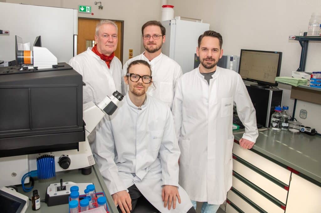
Researchers from the College Hospital Bonn (UKB) and the College of Bonn utilized high-performance microscopes to check the impact of various medicines on Staphylococcus aureus cell division.
They found that the manufacturing of peptidoglycan, a serious element of the bacterial cell wall, is the driving pressure for the entire cell division course of and that varied antibiotics block cell division in only a few minutes. Inside a couple of minutes, they found how totally different antibiotics block cell division.
The bacterial cell wall retains unicellular organisms in form and integrity, and cell wall synthesis is important for bacterial development.
The cell division protein FtsZ produces the so-called Z-ring within the heart of the cell, starting the division course of. A brand new cell wall is generated there, with peptidoglycan as the first element. On account of this constriction, two equivalent daughter cells are shaped.
The mannequin organism was chosen for investigation by the UKB analysis group led by Fabian Grein and Tanja Schneider and the group led by Ulrich Kubitscheck, Professor of Biophysical Chemistry. The examine targeted on the affect of antibiotics that inhibit peptidoglycan synthesis on cell division.
Jan-Samuel Puls, a Ph.D. pupil on the Institute of Pharmaceutical Microbiology at UKB, stated, “We discovered a fast and powerful impact of oxacillin and the glycopeptide antibiotics vancomycin and telavancin on cell division. The cell division protein FtsZ served as a marker right here, and we monitored it.”
FtsZ was fluorescently labeled alongside different proteins for that reason. The researchers subsequent utilized super-resolution microscopy to look at the consequences on particular person residing bacterial cells over time.

They developed an automatic picture evaluation system for microscope photos, permitting them to investigate all cells within the pattern underneath examine shortly. Dr. Fabian Grein is a scientist on the German Middle for An infection Analysis and a junior analysis group chief on the UKB’s Institute of Pharmaceutical Microbiology. (DZIF), clarify, “Staphylococcus aureus has a diameter of roughly one micrometer or one-thousandth of a millimeter. This makes microscopy significantly troublesome.”
The researchers from Bonn found that the formation of peptidoglycan is the driving pressure throughout the whole means of cell division. That inhibition of cell wall meeting by glycopeptide antibiotics in Staphylococcus aureus happens shortly and dramatically. Additionally they found the particular function of important penicillin-binding protein 2 (PBP2), which connects cell wall elements in cell division. The β-lactam antibiotic oxacillin prevents this protein from being correctly localized.
Grein says, “Because of this PBP2 doesn't get to the place the place it's wanted. Consequently, the cell can’t divide. Importantly, this all occurs instantly after the antibiotics are added. So the primary mobile results, which haven't been studied very intensively up to now, are essential.”
Due to the alarming enhance in antibiotic resistance worldwide, he hopes the examine outcomes will present a greater understanding of how precisely these brokers work on the mobile stage and thus a key to growing new antibiotics.
The outcomes of this examine will assist develop new antibiotics by offering a greater data of how these medicines work on the mobile stage.
Post a Comment